


line dot animation
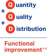
Functional improvement for those living with Duchenne requires dystrophin produced in suitable quantities, with sufficient quality, and distributed to the muscle cells most impacted by the disease.


Tripp, Age 6
Living with Duchenne
Understanding the foundational components of dystrophin that are critical to Duchenne pathophysiology
Duchenne is a progressive, multisystem disease with a definitive root cause: little to no functional dystrophin.7-10
What is dystrophin? i
Dystrophin is an essential structural protein found in all muscles of the human body, including skeletal, cardiac, and smooth muscle11-13*
- It is a key component of a large, multiprotein assembly known as the dystrophin‑associated protein complex (DAPC), localized to the sarcolemma, the membrane that surrounds the muscle cell14,15
- The DAPC stabilizes the sarcolemma and provides resilience during muscle contraction, allowing dystrophin to provide shock absorption2,14
Dystrophin links the intracellular actin cytoskeleton to the extracellular matrix through the DAPC, creating a structural scaffold across the sarcolemma2,16
*The structural and protective role of dystrophin and the DAPC in muscle has been extensively studied in striated muscle (skeletal and cardiac muscle). Its precise role in smooth muscle is not as well understood.11-13
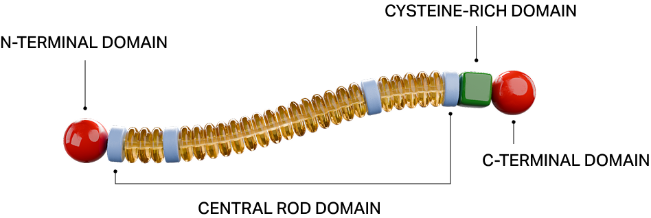
Specific domains within dystrophin interact with various cytoskeletal components for the protein to function appropriately2,17-19
The domains each play distinct structural roles that work in concert to stabilize the sarcolemma
How does dystrophin work? i
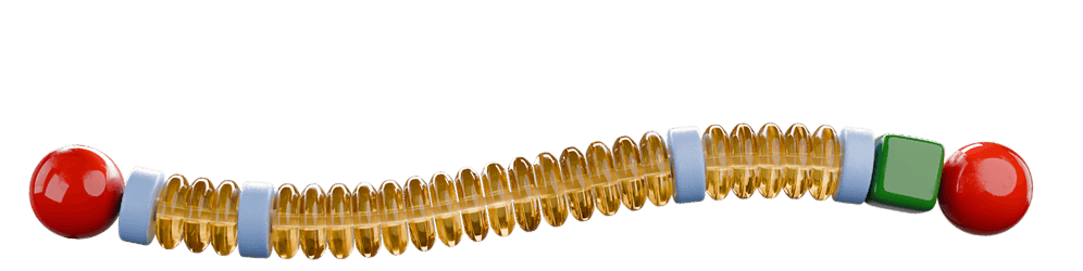
Image depicts the dystrophin protein.
How does dystrophin work? i
Image depicts the dystrophin protein.

Anchors dystrophin to the cytoskeleton and contractile apparatus
How does dystrophin work? i
Image depicts the dystrophin protein.
Composed of hinge regions and spectrin-like repeats, the central rod domain provides elasticity and flexibility, acting as a molecular shock absorber and helping protect the sarcolemma from damage
How does dystrophin work? i
Image depicts the dystrophin protein.
Facilitates binding to the sarcolemma, the membrane that surrounds the muscle cell
How does dystrophin work? i
Image depicts the dystrophin protein.
Mediates sarcolemma localization via the binding of dystrophin-associated proteins
What happens to skeletal, cardiac, and smooth muscle without functional dystrophin?i
The absence of functional dystrophin disrupts muscle integrity2,4
- In Duchenne, full-length functional dystrophin is produced at very low levels (or none at all), across all types of muscle and other tissues9,20,21
- This results in sarcolemmal fragility and increased permeability everywhere that functional dystrophin is very low or absent, leading to calcium dysregulation and free radical formation2
- The end result is muscle damage and degeneration, where damaged muscle cells are replaced by fat and fibrotic tissue that disrupts function in systems throughout the body2,4,7,22
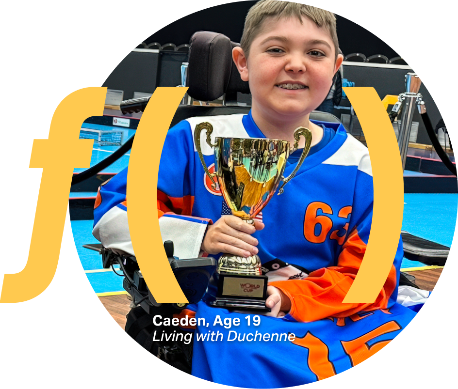
Caeden, Age 19
Living with Duchenne
Dystrophin restoration requires a biologic achievement of the 3 key components in the functional improvement equation: quantity, quality, and distribution.1-6
How does dystrophin quantity impact function? i
Increased dystrophin expression in Duchenne is associated with improved muscle biology, which may enhance structural muscle support and reduce muscle cell damage.23-28
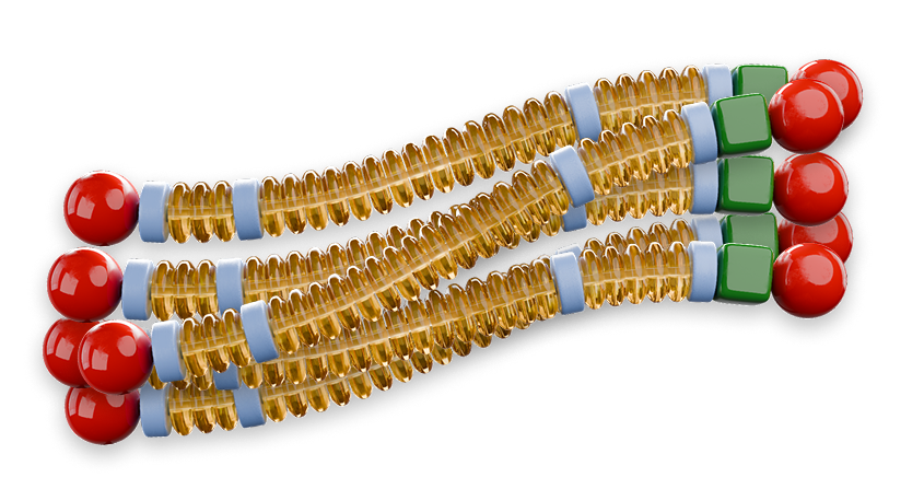
Preclinical studies and observational clinical studies have shown an association between higher dystrophin levels and delayed loss of ambulation and slower disease progression.23-28
- In studies comparing dystrophin levels and clinical phenotype, patients with very low levels of dystrophin were associated with more severe disease; those with higher levels had moderate or mild disease24,26,27
- Studies suggest that even low levels of residual dystrophin may provide functional benefit8,16,24,29,30
How does dystrophin quality impact function? i
For dystrophin to function properly, key domains critical to sarcolemmal stability and shock absorption must be expressed.2,16,31-34
Dystrophin restoration that is as close as possible to naturally occurring dystrophin is more likely to provide functional benefit and protect muscles.2,16,31-34
- Certain domains of dystrophin are critical. For example, the central actin-binding domain, ABD2, helps protect muscle from contraction-induced injury by bringing together the sarcolemma and actin filaments2,17,35
How does dystrophin distribution impact function? i
For meaningful functional improvement in Duchenne, dystrophin restoration needs to occur across all muscle types, and dystrophin should be appropriately distributed within muscle cells.1-4,8,16,22,36,37
Skeletal, cardiac, and smooth muscles have different structures and functions, but all express dystrophin; its role in sarcolemmal integrity has most directly been demonstrated in skeletal and cardiac muscle.2,3,38
- Skeletal muscle is composed of lengthy, multinucleated cells where it is thought that individual nuclei regulate the cytoplasmic area proximal to each one; these units are described as myonuclear domains (MNDs). Dystrophin restoration that does not extend beyond certain MNDs could still make the cell vulnerable to contraction-induced damage13,37,39-41
- Cardiac muscle is continuously active, and therefore has a different contractile phenotype compared to skeletal muscle. Cardiac muscle is less tolerant of heterogenous dystrophin expression and lacks the regenerative capacity present in skeletal muscle2,38,39,42-45
- Smooth muscle is widely distributed in the body, including the gastrointestinal (GI) tract and vasculature. Smooth muscle cells are organized and function differently based on tissue type, and it is believed that the role of dystrophin also varies; however, this has not been well studied8,38,46-48
Take a closer look at the science
Learn more about how each components plays a critical role in dystrophin production, and see how achieving them simultaneously could shift the goalpost in the treatment of Duchenne from slowing progression to delivering functional improvement.

Dystrophin-producing
approaches today
Advancements have been made that target the underlying cause of the disease, not just the symptoms.
Existing dystrophin-producing approaches have helped to establish awareness of the individual elements of dystrophin that are important for muscle function.4,49
What is exon skipping? i
- Exon skipping therapy uses an antisense oligonucleotide that binds pre-mRNA to alter the cell's machinery; this bypasses a specific exon during the splicing process, resulting in the production of functional, near full-length dystrophin50,51
- It was the first approach that proved near full-length dystrophin could be generated2,8,16,51,52
- Because of the way it is delivered, the therapy may not reach all the muscle cells impacted by Duchenne16,51,53
What is gene therapy? i
- Gene therapy involves packaging the micro-dystrophin transgene into an adeno-associated virus (AAV) vector that is targeted to muscle cells4
- The gene stays as a stable episome of the cell to produce robust expression of micro-dystrophin54,55
- Gene therapy is given once and may not be re-dosed. As the body naturally grows, new muscle cells impacted by Duchenne may not receive the medicine2,14,18
Current approaches slow, but do not stop disease progression8,14,16,50,56-58
Data show continued functional decline despite treatment.†

While there is a delay in the loss of ambulation, disease progression still occurs59

Cardiac and respiratory disease remains the leading drivers of morbidity and mortality in Duchenne60
†Corticosteroids, the standard of care, have been shown to delay disease progression. Exon skipping and gene therapy are newer approaches; however, data do not yet exist demonstrating ability to stop disease progression.
Achieving all 3 components of dystrophin simultaneously
remains one of the most persistent and defining challenges in the pursuit of functional improvement1-6
line dot animation

What’s next?
Hear from healthcare professionals, people living with Duchenne, and caregivers about the importance of creating a more functional future in Duchenne.

Tripp, Age 6
Living with Duchenne
REFERENCES:
1. Bengtsson NE, Tasfaout H, Chamberlain JS. Mol Ther. 2025;33(5):2035-2051. 2. Bez Batti Angulski A, Hosny N, Cohen H, et al. Front Physiol. 2023;14:1183101. 3. Elangkovan N, Dickson G. J Neuromuscul Dis. 2021;8(s2):S303-S316. 4. Loboda A, Chamberlain JS, Dulak J. Mol Ther Nucleic Acids. 2025;36(4):102759. 5. Takeda S, Clemens PR, Hoffman EP. J Neuromuscul Dis. 2021;8(s2):S343-S358. 6. Torelli S, Scaglioni D, Sardone V, et al. J Neuropathol Exp Neurol. 2021;80(10):955-965. 7. Duan D, Goemans N, Takeda S, Mercuri E, Aartsma-Rus A. Nat Rev Dis Primers. 2021;7(1):13. 8. Konieczny P. Clin Pharmacol Ther. 2024;116(6):1472-1484. 9. Leckie J, Zia A, Yokota T. Genes (Basel). 2024;15(11):1489. 10. Novelli V, Canonico F, Laborante R, et al. Biomedicines. 2025;13(1):102. 11. Aartsma-Rus A, Takeda S. J Neuromuscul Dis. 2025;13(2):147-155. 12. Kaplan KM, Morgan KG. Front Physiol. 2022;13:1059021. 13. Morin A, Stantzou A, Petrova ON, et al. Proc Natl Acad Sci U S A. 2023;120(2):e2206324120. 14. Kurshakova EV, Levchenko OA, Smirnikhina SA, Lavrov AV. Curr Issues Mol Biol. 2025;47(12):1058. 15. Tjondrokoesoemo A, Schips TG, Sargent MA, et al. J Biol Chem. 2016;291(19):9920-9928. 16. Roberts TC, Wood MJA, Davies KE. Nat Rev Drug Discov. 2023;22(11):917-934. 17. Gao QQ, McNally EM. Compr Physiol. 2015;5(3):1223-1239. 18. Hart CC, Lee YI, Xie J, et al. JCI Insight. 2024;9(11):e165869. 19. Szwec S, Kaplucha Z, Chamberlain JS, Konieczny P. BioDrugs. 2024;38(1):95-119. 20. García-Rodríguez R, Hiller M, Jiménez-Gracia L, et al. Proc Natl Acad Sci U S A. 2020;117(28):16456-16464. 21. Lu QL. J Neuromuscul Dis. 2021;8(s2):S359-S367. 22. Subhan F, Zizzo MG, Serio R. Neurogastroenterol Motil. 2024;36(8):e14804. 23. Birch SM, Lawlor MW, Conlon TJ, et al. Sci Transl Med. 2023;15(677):eabo1815. 24. de Feraudy Y, Ben Yaou R, Wahbi K, et al. Annals of Neurology. 2021;89(2):280-292. 25. Godfrey C, Muses S, McClorey G, et al. Hum Mol Genet. 2015;24(15):4225-4237. 26. Hoffman EP, Fischbeck KH, Brown RH, et al. N Engl J Med. 1988;318(21):1363-1368. 27. Hoffman EP, Kunkel LM, Angelini C, Clarke A, Johnson M, Harris JB. Neurology. 1989;39(8):1011-1017. 28. Le Guiner C, Servais L, Montus M, et al. Nat Commun. 2017;8:16105. 29. Etxaniz U, Marks I, Albin T, et al. Nucleic Acids Res. 2025;53(6). doi:10.1093/nar/gkaf241. 30. McDonald C, Camino E, Escandon R, et al. J Neuromuscul Dis. 2024;11(2):499-523. 31. Palmieri L, Ferrand M, Vu Hong A, Richard I, Albini S. Int J Mol Sci. 2024;25(19):10444. 32. Tasfaout H, Halbert CL, McMillen TS, et al. Nature. 2024;632(8023):192-200. 33. Torella A, Zanobio M, Zeuli R, et al. PLoS One. 2020;15(8):e0237803. 34. Waldrop MA, Yaou RB, Lucas KK, et al. J Neuromuscul Dis. 2020;7(3):217-229. 35. Mias-Lucquin D, Dos Santos Morais R, Chéron A, et al. J Struct Biol. 2020;209(1):107411. 36. Chwalenia K, Wood MJA, Roberts TC. J Muscle Res Cell Motil. 2025;46(4):293-300. 37. van Westering TLE, Lomonosova Y, Coenen-Stass AML, et al. J Cachexia Sarcopenia Muscle. 2020;11(2):578-593. 38. Sweeney HL, Hammers DW. Cold Spring Harb Perspect Biol. 2018;10(2):a023200. 39. Lu QL, Zhao S, Blaeser AR. Mol Ther. 2025;33(6):2517-2526. 40. Morandi L, Mora M, Confalonieri V, et al. J Neurol Sci. 1995;132(2):146-155. 41. Zhao J, Du X, Fang W, Zhu H, Yue B. Front Cell Dev Biol. 2025;13:1602607. 42. Bostick B, Yue Y, Long C, Duan D. Circ Res. 2008;102(1):121-130. 43. Meyers TA, Heitzman JA, Townsend D. Hum Mol Genet. 2020;29(6):944-954. 44. Przymuszala M, Bialobrzeska M, Dulak J, Florczyk-Soluch U. J Cachexia Sarcopenia Muscle. 2026;17(1):e70180. 45. Szedlak P, Steele DS, Hopkins PM. BJA Educ. 2023;23(9):350-357. 46. Carrington G, Zahedi M, Peckham M. J Muscle Res Cell Motil. 2026;47(2):13. 47. Sharma P, Basu S, Mitchell RW, Stelmack GL, Anderson JE, Halayko AJ. PLoS One. 2014;9(7):e102737. 48. Xuan W, Cheng F, Han X, Tipparaju SM, Ashraf M. Sci Rep. 2025;15(1):45254. 49. Roberts TC, Langer R, Wood MJA. Nat Rev Drug Discov. 2020;19(10):673-694. 50. Chassin A, Ono H, Ashida Y, Imamura M, Aoki Y. Int J Mol Sci. 2026;27(6):2755. 51. Owusu L, Kim S, Patel H, et al. Mol Ther Nucleic Acids. 2026;37(2):102901. 52. Molinari E, Srivastava S, Sayer JA, Ramsbottom SA. F1000Res. 2017;6:669. 53. Filonova G, Aartsma-Rus A. Expert Opin Biol Ther. 2023;23(2):133-143. 54. Mendell JR, Muntoni F, McDonald CM, et al. Nat Med. 2025;31(1):332-341. 55. Scoto M, Muntoni F. Med. 2025;6(12):100938. 56. Aartsma-Rus A, Jonker AH, O’Connor D. Neuromuscul Disord. 2025;55:106221. 57. Madaan P, Mishra SK, Wanigasinghe J, Sahu JK. Lancet Reg Health Southeast Asia. 2025;43:100699. 58. Pesco MT, Öztürk GZ, Bhadola SC, Chrzanowski SM, Gushchina LV, D’Ambrosio ES. Muscles. 2026;5(1):21. 59. PPMD. Parent Project Muscular Dystrophy. Fifteen Year Registry Report. 2023. Accessed May 13, 2026. https://www.parentprojectmd.org/wp-content/uploads/2023/08/PPMD_15-Year-Registry-Report_2023.pdf 60. Birnkrant DJ, Bushby K, Bann CM, et al. Lancet Neurol. 2018;17(5):445-455.
Tap the info button throughout the site to see a simplified summary of complex scientific topics
In Duchenne, the main cause is that the body does not make working dystrophin, the protein that provides a protective cushion for your muscles.
Dystrophin works like a shock absorber to help protect the muscles during contraction from everyday movements like walking, breathing, and the beating of the heart.
Without dystrophin, the muscles become fragile, causing them to be easily damaged and weaken over time with repeated use.
Dystrophin is a protein made up of several parts or domains that work together to help muscles function. Together, these different domains help dystrophin connect to important structures inside the muscle cell. Dystrophin cushions muscles during movement and helps stabilize the muscle cell’s protective barrier so it stays strong with everyday use.
In Duchenne, the body makes very little or no functional dystrophin. Without dystrophin, the protective barrier of the muscle cell becomes weaker, allowing too much calcium to enter and cause damage. Over time, the damaged muscle cells are replaced with fat and scar tissue.
Research shows that having more dystrophin can help muscles stay healthier in Duchenne. Higher levels of dystrophin are linked to a less severe disease course, which suggests muscles are better supported. People with higher levels of dystrophin continue to walk longer and experience slower weakening of muscle strength compared with those with lower levels of dystrophin.
There are several different muscle types—each with its own structure and function. Skeletal muscle is used for movement and breathing, cardiac muscle helps pump blood throughout the body, and smooth muscle is found in the digestive system. They all require dystrophin to work properly.
Duchenne affects the whole body, so for dystrophin to optimally impact function, it needs to be restored across all muscle types and within the muscle cells themselves. For example, restoring dystrophin only to some cardiac muscle cells or parts of the skeletal muscle cell could still lead to muscle instability and damage.
Exon skipping allows the body to skip over the exon next to the mutation restoring the connection so the body can make a shorter form of dystrophin. These medicines showed for the first time that it is possible to produce dystrophin that resembles the natural protein. Because of the way the medicine is delivered, it may not reach all of the muscle cells affected by Duchenne that need it most.
Gene therapy is designed to deliver "instructions" directly to muscle cells so they can make large amounts of micro-dystrophin, a shorter version of dystrophin. Gene therapy is given once and may not be re-dosed. As the body naturally grows, new muscle cells impacted by Duchenne may not receive the medicine.
The quality of dystrophin matters. Dystrophin that is close to what the body naturally makes can help protect muscles during movement and everyday use.